Yeast Kinetochore
In collaboration with Sue Biggins (FHCRC) we studied the structure of the yeast kinetochore by electron tomography. In collaboration with Trisha Davis and Chip Asbury (UW) we studied microtubule dynamics and microtubule binding proteins.

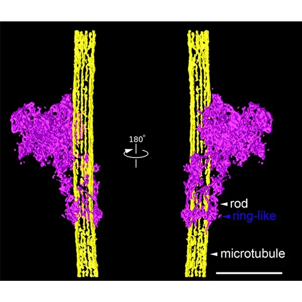
Chromosomes must be accurately partitioned to daughter cells to prevent genomic instability and aneuploidy, a hallmark of many tumors and birth defects. Kinetochores are macromolecular machines that move chromosomes by maintaining load-bearing attachments to the assembling and disassembling tips of spindle microtubules. The mechanism by which kinetochores attach to microtubules is still not clear although a number of models have been proposed. Sue’s laboratory previously developed an assay to purify functional native budding yeast kinetochore particles that contain the majority of core structural components and can maintain attachments to microtubules under force. We presented the structure of these isolated kinetochore particles as visualized by electron microscopy (EM) and electron tomography of negatively stained preparations. The budding yeast kinetochore appeared as a ~126 nm particle having a large central hub attached to multiple outer globular domains. Microtubule binding experiments indicated that the globular domains are important for microtubule attachments both in the presence or absence of a ring encircling the microtubule. Our data showed that kinetochores bind to microtubules via multivalent attachments, consistent with a biased diffusion mechanism where multiple kinetochore components cooperate to form a strong yet dynamic linkage to the microtubule. Although rings are not required for lateral binding, they likely maintain processive attachments to the ends of dynamic microtubules. These studies lay the foundation to uncover the key mechanical and regulatory mechanisms by which kinetochores control chromosome segregation and cell division.
Relevant papers
Umbreit, Neil T.; Gestaut, Daniel R.; Tien, Jerry F.; Vollmar, Breanna S.; Gonen, Tamir; Asbury, Charles L.; Davis, Trisha N.
The Ndc80 kinetochore complex directly modulates microtubule dynamics
In: Proc. Natl. Acad. Sci. U.S.A., vol. 109, no. 40, pp. 16113–16118, 2012.
Gonen, Shane; Akiyoshi, Bungo; Iadanza, Matthew G; Shi, Dan; Duggan, Nicole; Biggins, Sue; Gonen, Tamir
The structure of purified kinetochores reveals multiple microtubule-attachment sites
In: Nat. Struct. Mol. Biol., vol. 19, no. 9, pp. 925–929, 2012.
Akiyoshi, Bungo; Sarangapani, Krishna K.; Powers, Andrew F.; Nelson, Christian R.; Reichow, Steve L.; Arellano-Santoyo, Hugo; Gonen, Tamir; Ranish, Jeffrey A.; Asbury, Charles L.; Biggins, Sue
Tension directly stabilizes reconstituted kinetochore-microtubule attachments
In: Nature, vol. 468, no. 7323, pp. 576–579, 2010.
Tien, Jerry F; Umbreit, Neil T; Gestaut, Daniel R; Franck, Andrew D; Cooper, Jeremy; Wordeman, Linda; Gonen, Tamir; Asbury, Charles L; Davis, Trisha N
Cooperation of the Dam1 and Ndc80 kinetochore complexes enhances microtubule coupling and is regulated by aurora B
In: J. Cell Biol., vol. 189, no. 4, pp. 713–723, 2010.
Franck, Andrew D; Powers, Andrew F; Gestaut, Daniel R; Gonen, Tamir; Davis, Trisha N; Asbury, Charles L
Tension applied through the Dam1 complex promotes microtubule elongation providing a direct mechanism for length control in mitosis
In: Nat. Cell Biol., vol. 9, no. 7, pp. 832–837, 2007.