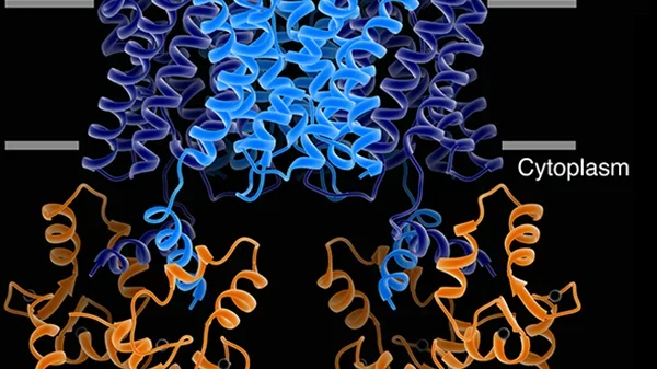
Our structure of the AQP0/CaM complex is the first for any full-length membrane channel in complex with this ubiquitous secondary messenger. Current efforts in the laboratory are to understand how Ca²⁺/CaM binds to and modulates the activity of other channels such as ion channels.
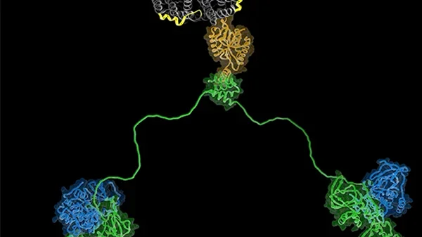
We are also trying to understand more about the AQP-AKAP system, in particular we are trying to assemble the AQP2-AKAP18-PKA complex and AQP0-AKAP2-PKA complex for structural studies. Intrinsically disordered regions of proteins are widespread in nature yet the mechanistic roles they play in biology are underappreciated. Such disordered segments can act simply to link functionally coupled structural domains or they can orchestrate enzymatic reactions through a variety of allosteric mechanisms. The regulatory subunits of protein kinase A provide an example of this important phenomenon where functionally defined and structurally conserved domains are connected by intrinsically disordered regions of defined length with limited sequence identity. Our studies show that this seemingly paradoxical amalgam of order and disorder permits fine-tuning of local protein phosphorylation events. The anchoring of PKA by AKAP affords the kinase a sphere of action in which multiple targets can get phosphorylated fast in a cAMP independent way.
Relevant papers
Smith, Donelson F; Reichow, Steve L; Esseltine, Jessica L; Shi, Dan; Langeberg, Lorene K; Scott, John D; Gonen, Tamir
Intrinsic disorder within an AKAP-protein kinase A complex guides local substrate phosphorylation
In: Elife, vol. 2, pp. e01319, 2013.
@article{pmid24192038,
title = {Intrinsic disorder within an AKAP-protein kinase A complex guides local substrate phosphorylation},
author = {Donelson F Smith and Steve L Reichow and Jessica L Esseltine and Dan Shi and Lorene K Langeberg and John D Scott and Tamir Gonen},
url = {https://cryoem.ucla.edu/wp-content/uploads/2013_smith.pdf, Main text},
doi = {10.7554/eLife.01319},
year = {2013},
date = {2013-11-05},
journal = {Elife},
volume = {2},
pages = {e01319},
abstract = {Anchoring proteins sequester kinases with their substrates to locally disseminate intracellular signals and avert indiscriminate transmission of these responses throughout the cell. Mechanistic understanding of this process is hampered by limited structural information on these macromolecular complexes. A-kinase anchoring proteins (AKAPs) spatially constrain phosphorylation by cAMP-dependent protein kinases (PKA). Electron microscopy and three-dimensional reconstructions of type-II PKA-AKAP18γ complexes reveal hetero-pentameric assemblies that adopt a range of flexible tripartite configurations. Intrinsically disordered regions within each PKA regulatory subunit impart the molecular plasticity that affords an ∼16 nanometer radius of motion to the associated catalytic subunits. Manipulating flexibility within the PKA holoenzyme augmented basal and cAMP responsive phosphorylation of AKAP-associated substrates. Cell-based analyses suggest that the catalytic subunit remains within type-II PKA-AKAP18γ complexes upon cAMP elevation. We propose that the dynamic movement of kinase sub-structures, in concert with the static AKAP-regulatory subunit interface, generates a solid-state signaling microenvironment for substrate phosphorylation. DOI: http://dx.doi.org/10.7554/eLife.01319.001.},
keywords = {},
pubstate = {published},
tppubtype = {article}
}
Reichow, Steve L; Clemens, Daniel M; Freites, Alfredo J; Nemeth-Cahalan, Karin L; Heyden, Matthias; Tobias, Douglas J; Hall, James E; Gonen, Tamir
Allosteric mechanism of water-channel gating by Ca²⁺-calmodulin
In: Nat. Struct. Mol. Biol., vol. 20, no. 9, pp. 1085–1092, 2013.
@article{pmid23893133,
title = {Allosteric mechanism of water-channel gating by Ca²⁺-calmodulin},
author = {Steve L Reichow and Daniel M Clemens and Alfredo J Freites and Karin L Nemeth-Cahalan and Matthias Heyden and Douglas J Tobias and James E Hall and Tamir Gonen},
url = {https://cryoem.ucla.edu/wp-content/uploads/2013_reichow.pdf, Main text},
doi = {10.1038/nsmb.2630},
year = {2013},
date = {2013-07-28},
journal = {Nat. Struct. Mol. Biol.},
volume = {20},
number = {9},
pages = {1085--1092},
abstract = {Calmodulin (CaM) is a universal regulatory protein that communicates the presence of calcium to its molecular targets and correspondingly modulates their function. This key signaling protein is important for controlling the activity of hundreds of membrane channels and transporters. However, understanding of the structural mechanisms driving CaM regulation of full-length membrane proteins has remained elusive. In this study, we determined the pseudoatomic structure of full-length mammalian aquaporin-0 (AQP0, Bos taurus) in complex with CaM, using EM to elucidate how this signaling protein modulates water-channel function. Molecular dynamics and functional mutation studies reveal how CaM binding inhibits AQP0 water permeability by allosterically closing the cytoplasmic gate of AQP0. Our mechanistic model provides new insight, only possible in the context of the fully assembled channel, into how CaM regulates multimeric channels by facilitating cooperativity between adjacent subunits.},
keywords = {},
pubstate = {published},
tppubtype = {article}
}
Gold, Matthew G; Reichow, Steve L; O'Neill, Susan E; Weisbrod, Chad R; Langeberg, Lorene K; Bruce, James E; Gonen, Tamir; Scott, John D
AKAP2 anchors PKA with aquaporin-0 to support ocular lens transparency
In: EMBO Mol Med, vol. 4, no. 1, pp. 15–26, 2011.
@article{pmid22095752,
title = {AKAP2 anchors PKA with aquaporin-0 to support ocular lens transparency},
author = {Matthew G Gold and Steve L Reichow and Susan E O'Neill and Chad R Weisbrod and Lorene K Langeberg and James E Bruce and Tamir Gonen and John D Scott},
url = {https://cryoem.ucla.edu/wp-content/uploads/2012_gold.pdf, Main text},
doi = {10.1002/emmm.201100184},
year = {2011},
date = {2011-11-16},
journal = {EMBO Mol Med},
volume = {4},
number = {1},
pages = {15--26},
abstract = {A decline in ocular lens transparency known as cataract afflicts 90% of individuals by the age 70. Chronic deterioration of lens tissue occurs as a pathophysiological consequence of defective water and nutrient circulation through channel and transporter proteins. A key component is the aquaporin-0 (AQP0) water channel whose permeability is tightly regulated in healthy lenses. Using a variety of cellular and biochemical approaches we have discovered that products of the A-kinase anchoring protein 2 gene (AKAP2/AKAP-KL) form a stable complex with AQP0 to sequester protein kinase A (PKA) with the channel. This permits PKA phosphorylation of serine 235 within a calmodulin (CaM)-binding domain of AQP0. The additional negative charge introduced by phosphoserine 235 perturbs electrostatic interactions between AQP0 and CaM to favour water influx through the channel. In isolated mouse lenses, displacement of PKA from the AKAP2-AQP0 channel complex promotes cortical cataracts as characterized by severe opacities and cellular damage. Thus, anchored PKA modulation of AQP0 is a homeostatic mechanism that must be physically intact to preserve lens transparency.},
keywords = {},
pubstate = {published},
tppubtype = {article}
}
Reichow, Steve L; Gonen, Tamir
Noncanonical Binding of Calmodulin to Aquaporin-0: Implications for Channel Regulation
In: Structure, vol. 16, no. 9, pp. 1389–1398, 2008.
@article{pmid18786401,
title = {Noncanonical Binding of Calmodulin to Aquaporin-0: Implications for Channel Regulation},
author = {Steve L Reichow and Tamir Gonen},
url = {https://cryoem.ucla.edu/wp-content/uploads/reichow_2008.pdf, Main text},
doi = {10.1016/j.str.2008.06.011},
year = {2008},
date = {2008-09-10},
journal = {Structure},
volume = {16},
number = {9},
pages = {1389--1398},
abstract = {Aquaporins (AQPs) are a family of ubiquitous membrane channels that conduct water across cell membranes. AQPs form homotetramers containing four functional and independent water pores. Aquaporin-0 (AQP0) is expressed in the eye lens, where its water permeability is regulated by calmodulin (CaM). Here we use a combination of biochemical methods and NMR spectroscopy to probe the interaction between AQP0 and CaM. We show that CaM binds the AQP0 C-terminal domain in a calcium-dependent manner. We demonstrate that only two CaM molecules bind a single AQP0 tetramer in a noncanonical fashion, suggesting a form of cooperativity between AQP0 monomers. Based on these results, we derive a structural model of the AQP0/CaM complex, which suggests CaM may be inhibitory to channel permeability by capping the vestibules of two monomers within the AQP0 tetramer. Finally, phosphorylation within AQP0's CaM binding domain inhibits the AQP0/CaM interaction, suggesting a temporal regulatory mechanism for complex formation.},
keywords = {},
pubstate = {published},
tppubtype = {article}
}
Gonen, Tamir; Cheng, Yifan; Sliz, Piotr; Hiroaki, Yoko; Fujiyoshi, Yoshinori; Harrison, Stephen C; Walz, Thomas
Lipid-protein interactions in double-layered two-dimensional AQP0 crystals
In: Nature, vol. 438, no. 7068, pp. 633–638, 2005.
@article{pmid16319884,
title = {Lipid-protein interactions in double-layered two-dimensional AQP0 crystals},
author = {Tamir Gonen and Yifan Cheng and Piotr Sliz and Yoko Hiroaki and Yoshinori Fujiyoshi and Stephen C Harrison and Thomas Walz},
url = {https://cryoem.ucla.edu/wp-content/uploads/Gonen_2005.pdf, Main text},
doi = {10.1038/nature04321},
year = {2005},
date = {2005-12-01},
journal = {Nature},
volume = {438},
number = {7068},
pages = {633--638},
abstract = {Lens-specific aquaporin-0 (AQP0) functions as a specific water pore and forms the thin junctions between fibre cells. Here we describe a 1.9 A resolution structure of junctional AQP0, determined by electron crystallography of double-layered two-dimensional crystals. Comparison of junctional and non-junctional AQP0 structures shows that junction formation depends on a conformational switch in an extracellular loop, which may result from cleavage of the cytoplasmic amino and carboxy termini. In the centre of the water pathway, the closed pore in junctional AQP0 retains only three water molecules, which are too widely spaced to form hydrogen bonds with each other. Packing interactions between AQP0 tetramers in the crystalline array are mediated by lipid molecules, which assume preferred conformations. We were therefore able to build an atomic model for the lipid bilayer surrounding the AQP0 tetramers, and we describe lipid-protein interactions.},
keywords = {},
pubstate = {published},
tppubtype = {article}
}
Gonen, Tamir; Cheng, Yifan; Kistler, Joerg; Walz, Thomas
Aquaporin-0 Membrane Junctions Form Upon Proteolytic Cleavage
In: J. Mol. Biol., vol. 342, no. 4, pp. 1337–1345, 2004.
@article{pmid15351655,
title = {Aquaporin-0 Membrane Junctions Form Upon Proteolytic Cleavage},
author = {Tamir Gonen and Yifan Cheng and Joerg Kistler and Thomas Walz},
url = {https://cryoem.ucla.edu/wp-content/uploads/Gonen_2004b.pdf, Main text},
doi = {10.1016/j.jmb.2004.07.076},
year = {2004},
date = {2004-09-24},
journal = {J. Mol. Biol.},
volume = {342},
number = {4},
pages = {1337--1345},
abstract = {Aquaporin-0 (AQP0), previously known as major intrinsic protein (MIP), is the only water pore protein expressed in lens fiber cells. AQP0 is highly specific to lens fiber cells and constitutes the most abundant intrinsic membrane protein in these cells. The protein is initially expressed as a full-length protein in young fiber cells in the lens cortex, but becomes increasingly cleaved in the lens core region. Reconstitution of AQP0 isolated from the core of sheep lenses containing a proportion of truncated protein, produced double-layered two-dimensional (2D) crystals, which displayed the same dimensions as the thin 11 nm lens fiber cell junctions, which are prominent in the lens core. In contrast reconstitution of full-length AQP0 isolated from the lens cortex reproducibly yielded single-layered 2D crystals. We present electron diffraction patterns and projection maps of both crystal types. We show that cleavage of the intracellular C terminus enhances the adhesive properties of the extracellular surface of AQP0, indicating a conformational change in the molecule. This change of function of AQP0 from a water pore in the cortex to an adhesion molecule in the lens core constitutes another manifestation of the gene sharing concept originally proposed on the basis of the dual function of crystallins.},
keywords = {},
pubstate = {published},
tppubtype = {article}
}
Gonen, Tamir; Sliz, Piotr; Kistler, Joerg; Cheng, Yifan; Walz, Thomas
Aquaporin-0 membrane junctions reveal the structure of a closed water pore
In: Nature, vol. 429, no. 6988, pp. 193–197, 2004.
@article{pmid15141214,
title = {Aquaporin-0 membrane junctions reveal the structure of a closed water pore},
author = {Tamir Gonen and Piotr Sliz and Joerg Kistler and Yifan Cheng and Thomas Walz},
url = {https://cryoem.ucla.edu/wp-content/uploads/gonen_2004a.pdf, Main text},
doi = {10.1038/nature02503},
year = {2004},
date = {2004-05-13},
journal = {Nature},
volume = {429},
number = {6988},
pages = {193--197},
abstract = {The lens-specific water pore aquaporin-0 (AQP0) is the only aquaporin known to form membrane junctions in vivo. We show here that AQP0 from the lens core, containing some carboxy-terminally cleaved AQP0, forms double-layered crystals that recapitulate in vivo junctions. We present the structure of the AQP0 membrane junction as determined by electron crystallography. The junction is formed by three localized interactions between AQP0 molecules in adjoining membranes, mainly mediated by proline residues conserved in AQP0s from different species but not present in most other aquaporins. Whereas all previously determined aquaporin structures show the pore in an open conformation, the water pore is closed in AQP0 junctions. The water pathway in AQP0 also contains an additional pore constriction, not seen in other known aquaporin structures, which may be responsible for pore gating.},
keywords = {},
pubstate = {published},
tppubtype = {article}
}
Relevant reviews
Gold, Matthew G; Gonen, Tamir; Scott, John D
Local cAMP signaling in disease at a glance
In: J. Cell. Sci., vol. 126, no. Pt 20, pp. 4537–4543, 2013.
@article{pmid24124191,
title = {Local cAMP signaling in disease at a glance},
author = {Matthew G Gold and Tamir Gonen and John D Scott},
url = {https://cryoem.ucla.edu/wp-content/uploads/2013_gold.pdf, Main text},
doi = {10.1242/jcs.133751},
year = {2013},
date = {2013-10-15},
journal = {J. Cell. Sci.},
volume = {126},
number = {Pt 20},
pages = {4537--4543},
abstract = {The second messenger cyclic AMP (cAMP) operates in discrete subcellular regions within which proteins that synthesize, break down or respond to the second messenger are precisely organized. A burgeoning knowledge of compartmentalized cAMP signaling is revealing how the local control of signaling enzyme activity impacts upon disease. The aim of this Cell Science at a Glance article and the accompanying poster is to highlight how misregulation of local cyclic AMP signaling can have pathophysiological consequences. We first introduce the core molecular machinery for cAMP signaling, which includes the cAMP-dependent protein kinase (PKA), and then consider the role of A-kinase anchoring proteins (AKAPs) in coordinating different cAMP-responsive proteins. The latter sections illustrate the emerging role of local cAMP signaling in four disease areas: cataracts, cancer, diabetes and cardiovascular diseases.},
keywords = {},
pubstate = {published},
tppubtype = {article}
}
Reichow, Steve L; Gonen, Tamir
Lipid-protein interactions probed by electron crystallography
In: Curr. Opin. Struct. Biol., vol. 19, no. 5, pp. 560–565, 2009.
@article{pmid19679462,
title = {Lipid-protein interactions probed by electron crystallography},
author = {Steve L Reichow and Tamir Gonen},
url = {https://cryoem.ucla.edu/wp-content/uploads/reichowgonen_2009.pdf, Main text},
doi = {10.1016/j.sbi.2009.07.012},
year = {2009},
date = {2009-10-01},
journal = {Curr. Opin. Struct. Biol.},
volume = {19},
number = {5},
pages = {560--565},
abstract = {Electron crystallography is arguably the only electron cryomicroscopy (cryoEM) technique able to deliver an atomic-resolution structure of membrane proteins embedded in the lipid bilayer. In the electron crystallographic structures of the light driven ion pump, bacteriorhodopsin, and the water channel, aquaporin-0, sufficiently high resolution was obtained and both lipid and protein were visualized, modeled, and described in detail. An extensive network of lipid-protein interactions mimicking native membranes is established and maintained in two-dimensional (2D) crystalline vesicles used for structural analysis by electron crystallography. Lipids are tightly integrated into the protein's architecture where they can affect the function, structure, quaternary assembly, and the stability of the membrane protein.},
keywords = {},
pubstate = {published},
tppubtype = {article}
}
Andrews, Simeon; Reichow, Steve L.; Gonen, Tamir
Electron Crystallography of Aquaporins
In: IUBMB Life, vol. 60, no. 7, pp. 430–436, 2008.
@article{pmid18465794,
title = {Electron Crystallography of Aquaporins},
author = {Andrews, Simeon and Reichow, Steve L. and Gonen, Tamir},
url = {https://cryoem.ucla.edu/wp-content/uploads/andrews_2008.pdf, Main text},
doi = {10.1002/iub.53},
year = {2008},
date = {2008-05-08},
journal = {IUBMB Life},
volume = {60},
number = {7},
pages = {430--436},
abstract = {Aquaporins are a family of ubiquitous membrane proteins that form a pore for the permeation of water. Both electron and X-ray crystallography played major roles in determining the atomic structures of a number of aquaporins. This review focuses on electron crystallography, and its contribution to the field of aquaporin biology. We briefly discuss electron crystallography and the two-dimensional crystallization process. We describe features of aquaporins common to both electron and X-ray crystallographic structures; as well as some structural insights unique to electron crystallography, including aquaporin junction formation and lipid-protein interactions.},
keywords = {},
pubstate = {published},
tppubtype = {article}
}
Engel, Andreas; Fujiyoshi, Yoshinori; Gonen, Tamir; Walz, Thomas
In: Curr. Opin. Struct. Biol., vol. 18, no. 2, pp. 229–235, 2008.
@article{pmid18194855,
title = {Junction-forming aquaporins},
author = {Andreas Engel and Yoshinori Fujiyoshi and Tamir Gonen and Thomas Walz},
url = {https://cryoem.ucla.edu/wp-content/uploads/engel_2008.pdf, Main text},
doi = {10.1016/j.sbi.2007.11.003},
year = {2008},
date = {2008-04-01},
journal = {Curr. Opin. Struct. Biol.},
volume = {18},
number = {2},
pages = {229--235},
abstract = {Aquaporins (AQPs) are a family of ubiquitous membrane channels that conduct water and solutes across membranes. This review focuses on AQP0 and AQP4, which in addition to forming water channels also appear to play a role in cell adhesion. We discuss the recently determined structures of the membrane junctions mediated by these two AQPs, the mechanisms that regulate junction formation, and evidence that supports a role for AQP0 and AQP4 in cell adhesion.},
keywords = {},
pubstate = {published},
tppubtype = {article}
}
Gonen, Tamir; Walz, Thomas
In: Q. Rev. Biophys., vol. 39, no. 4, pp. 361–396, 2006.
@article{pmid17156589,
title = {The structure of aquaporins},
author = {Tamir Gonen and Thomas Walz},
url = {https://cryoem.ucla.edu/wp-content/uploads/gonenwalz_2006.pdf, Main text},
doi = {10.1017/S0033583506004458},
year = {2006},
date = {2006-11-01},
journal = {Q. Rev. Biophys.},
volume = {39},
number = {4},
pages = {361--396},
abstract = {The ubiquitous members of the aquaporin (AQP) family form transmembrane pores that are either exclusive for water (aquaporins) or are also permeable for other small neutral solutes such as glycerol (aquaglyceroporins). The purpose of this review is to provide an overview of our current knowledge of AQP structures and to describe the structural features that define the function of these membrane pores. The review will discuss the mechanisms governing water conduction, proton exclusion and substrate specificity, and how the pore permeability is regulated in different members of the AQP family.},
keywords = {},
pubstate = {published},
tppubtype = {article}
}