Fragment‐Based Phase Extension
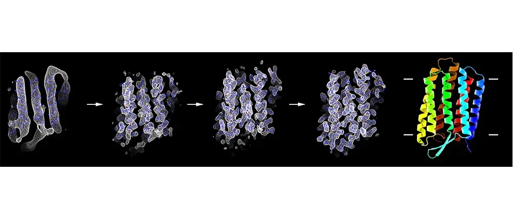
In electron crystallography membrane protein structure is determined from two-dimensional crystals where the protein is embedded in a membrane. Once large and well-ordered 2D crystals are grown one of the bottlenecks in electron crystallography is the collection of image data to directly provide experimental phases to high resolution. We developed a new approach to bypass this bottleneck, eliminating the need for high-resolution imaging. We used the strengths of electron crystallography in rapidly obtaining accurate experimental phase information from low-resolution images and accurate high-resolution amplitude information from electron diffraction. The low-resolution experimental phases were used for the placement of α-helix fragments and extended to high resolution using phases from the fragments. Phases were further improved by density modifications followed by fragment expansion and structure refinement against the high-resolution diffraction data. Using this approach, structures of three membrane proteins were determined rapidly and accurately to atomic resolution without high-resolution image data.
Relevant Papers
Nguyen, Chi; Lei, Hsiang-Ting; Lai, Louis Tung Faat; Gallenito, Marc J.; Matthies, Doreen; Gonen, Tamir
Lipid flipping in the omega-3 fatty-acid transporter
In: bioRxiv 2022, visited: 01.06.2022.